Traumatic brain injury leaves many victims emotionally shattered and cognitively crippled. But because mild and moderate brain injuries do not show up on CT or other imaging, doctors and even family members are often skeptical that any real damage exists. This skepticism can exacerbate the feelings of isolation and frustration that many victims experience, as they struggle to convey their pain and challenges. Recent traumatic brain injury research findings highlight the profound impact that even mild concussions can have on mental health and cognitive functioning over time. As awareness of these hidden injuries grows, it becomes increasingly important for both medical professionals and families to recognize and validate the experiences of those affected.
Now the first experiment of its kind documents exactly what “the invisible injury” – at least the kind caused by blast waves or physical impacts – does to the brain: Crumpled axons, which carry signals between neurons; gummed-up neurons like those in Alzheimer’s disease; strangled blood vessels.
A new study, published this month in the journal Science Translational Medicine, compared three groups of brains that had sustained concussions — young adults from military and athletic arenas, as well as animal brains.
The damage identified in the study was strikingly similar to what scientists have seen in the brains of ex-football players who had sustained head injuries and, after death, were found to have chronic traumatic encephalopathy (CTE), the condition once known as boxer’s dementia.
CTE can cause depression, aggression, impulsivity and memory loss and has been linked to suicide.
The results of the study also suggest that head trauma should be treated immediately instead of waiting for symptoms. The U.S. military has a policy of identifying TBI as soon as possible. That policy applies regardless of whether the injury is obvious and severe, or subtle and hidden. All service members in a vehicle collision or rollover or within 150 feet of a blast undergo a mandatory medical evaluation.
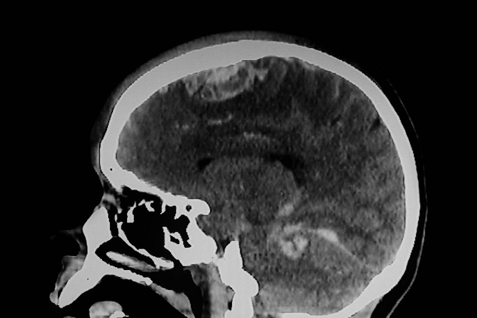
The study also confirms the long-standing notion that CT and MRI lack the resolution to show the cellular and sub-cellular changes that occur from a concussion or sub-concussive injury.
Abstract of the study can be reviewed here: http://stm.sciencemag.org/content/4/134/134ra60.abstract.